High-Dose-Rate (HDR) Brachytherapy with Remote Afterloading
Keywords: remote afterloading, brachytherapy, radioactive source implantation, three-dimensional planning, intracavitary irradiation, local dose escalation
Abstract: Remote afterloading brachytherapy is a form of brachytherapy in which a radioactive source is placed in close proximity to the tumor to deliver localized irradiation, achieving high-dose, targeted tumor control. It is commonly combined with external beam radiotherapy and is suitable for definitive treatment or local dose escalation in intracavitary and superficial tumors, such as cervical and esophageal cancers.
Applicable Situations
① Typical scenarios particularly suitable for remote afterloading brachytherapy
• Accessible location: The lesion is located in a natural cavity or on a superficial/mucosal surface (such as the cervix, vagina, esophagus, bronchus, rectum, skin, oral cavity), allowing applicators to be placed close to the target area.
• Need for local dose escalation: The lesion is adjacent to critical organs, and dose needs to be concentrated on the target while reducing irradiation to surrounding tissues.
• Boost after external beam radiotherapy: Additional local dose escalation to the primary lesion or residual disease after completion of external beam radiotherapy.
• Postoperative adjuvant therapy: Based on pathological findings and recurrence risk assessment, additional irradiation to local high-risk areas is required.
• Symptom relief: Tumor-related bleeding or luminal stenosis/obstruction, where brachytherapy may be used for hemostasis or symptom palliation after evaluation.
② Common indications
• Intracavitary treatment: lung cancer, esophageal cancer, rectal cancer, cervical cancer, nasopharyngeal cancer
• Interstitial implantation therapy: superficial solid tumors, bone tumors, breast cancer, prostate cancer, tongue cancer
• Intraoperative implantation with postoperative treatment: cholangiocarcinoma, pancreatic cancer, liver cancer, intra-abdominal masses
• Surface applicator brachytherapy: skin cancer, oral cancer
The specific suitability, treatment modality, and course schedule should be determined by the radiotherapy team after comprehensive evaluation based on imaging and examination results.
Technical Advantages
① Closer to the target, suitable for local dose escalation
The radioactive source can be delivered via the applicator into the tumor cavity or adjacent tissue, allowing it to be closer to the target area. External beam paths must pass through normal tissue, limiting local dose escalation. When appropriate, HDR brachytherapy is more favorable for dose intensification in the tumor core.
② More concentrated dose, better control over surrounding organs
The source can be delivered through a catheter to the vicinity or inside the tumor, concentrating the dose on the area needing reinforcement. The dose drops rapidly outside the target area. For lesions near critical organs like the bladder or rectum, this helps ensure the tumor receives sufficient dose while minimizing exposure to surrounding tissues.
③ Combination of intracavitary and external irradiation for comprehensive treatment
Because of its localized concentration and minimal exposure to surrounding tissue, many clinical cases adopt a combination of intracavitary and external beam therapy (e.g., cervical cancer, endometrial cancer). External beams cover the overall treatment area, while intracavitary therapy reinforces the primary lesion or high-risk regions, providing more complete and targeted treatment.
④ 3D planning and interstitial techniques for individualized plans
Compared with traditional 2D methods, 3D HDR planning allows for precise dose design and evaluation based on imaging, and applicators can be selected according to tumor extent and anatomy. When intracavitary coverage is insufficient, interstitial implantation can be combined to optimize dose distribution according to the tumor volume.
Why Choose Jinshazhou Hospital?
In afterloading brachytherapy, the quality of treatment and patient experience is determined not only by equipment but also by 3D planning capability, team coordination, integration with external beam radiotherapy, and full-process quality control. Our hospital has comprehensive configuration and experience in these critical areas.
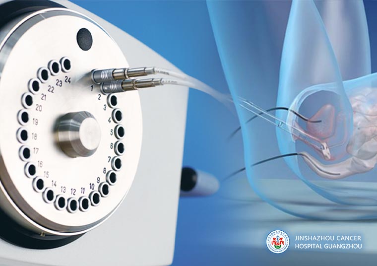
Our hospital is equipped with the Varian GammaMedplus iX high-dose-rate (HDR) brachytherapy system, which allows 3D planning and dose evaluation based on imaging localization, and enables individualized optimization under organ dose constraints. The system supports remote automatic source delivery and retrieval, and is compatible with multiple types of intracavitary applicators, facilitating adaptation to different anatomical structures and treatment requirements, making plan execution more stable and controllable.
At our hospital, afterloading brachytherapy is usually not an isolated procedure but part of an integrated treatment approach. Through MDT collaboration, we can coordinate brachytherapy smoothly with external beam radiotherapy, and when necessary, interventional, minimally invasive, or systemic treatments. Our external beam radiotherapy platform is fully equipped with flexible device and technical pathway options, allowing combined strategies of external beam coverage and brachytherapy boost, forming a complete one-stop treatment process.
Regarding safety and patient experience, we have established quality control and safety management procedures, standardizing equipment status checks, plan review, critical step verification, patient positioning, and treatment delivery, while providing post-treatment follow-up and reaction management recommendations.
For patients, this means a more stable treatment process, clearer care pathway, and greater attention to comfort and post-treatment management. For international patients, we provide multilingual communication support and assistance with treatment arrangements, ensuring smoother evaluation, therapy, and follow-up.
Technical Definition
Afterloading brachytherapy, also referred to as close-distance radiotherapy / intracavitary irradiation or sealed-source therapy, involves first placing an applicator or catheter without the radioactive source into the appropriate position; the device then delivers the radioactive source through the channel according to the treatment plan, and the source is automatically retracted after the irradiation is completed---hence the term afterloading. This technique can be used independently, but it is often combined with surgery, external beam radiotherapy, and necessary systemic therapy to achieve a complementary effect.
Mechanism of Action / Working Principle
① Automated source delivery irradiation: During treatment, the applicator or catheter is first positioned. During the irradiation phase, the afterloading device delivers the radioactive source along the catheter to pre-planned dwell positions under shielding and remote control. The source remains at each dwell point for the prescribed time and is automatically retracted after irradiation, so no radioactive material remains in the body.
② Effect of radiation on the tumor: The radiation released by the source acts on tumor cells, causing DNA damage directly or indirectly through free radicals, inhibiting tumor cell growth and division, thereby achieving the therapeutic goal. Since the source is close to the lesion, the target area receives a more concentrated dose, which decreases rapidly outside the target.
③ High Dose Rate (HDR): HDR refers to a higher dose delivered per unit time, so the actual single irradiation session is usually shorter (often measured in minutes). This makes the irradiation process more efficient, reduces the time the patient needs to maintain a fixed position, and improves the overall treatment experience.
Treatment Procedure
① Evaluation, examination, and treatment planning discussion: Assess suitability for afterloading brachytherapy through imaging studies and necessary blood tests. The doctor explains the treatment method, precautions, and whether combined external beam radiotherapy is needed, and completes the informed consent process.
② Pre-treatment preparation: Follow medical instructions for diet and medication adjustments; on the day of treatment, empty bladder and bowels as required. In some cases, bladder filling or holding urine may be necessary.
③ Applicator or catheter placement: Under local anesthesia, sedation, or general anesthesia, the applicator is placed via a natural cavity, or interstitial catheters/needles are inserted as needed.
④ Imaging localization and treatment plan confirmation: Perform CT/MRI localization; create a 3D plan and evaluate doses based on imaging; confirm irradiation positions and dwell times.
⑤ Irradiation delivery and post-treatment observation: The afterloading device delivers the source remotely and automatically; after irradiation, the source is retracted and applicators removed (or handled according to medical instructions). Post-treatment monitoring includes checking for bleeding, pain, infection, and providing nursing care and follow-up arrangements.
Frequently Asked Questions
① Will the treatment hurt? Is anesthesia needed?
Answer: Discomfort mostly occurs during the placement of the applicator or catheter and varies by individual. The doctor may provide local anesthesia, sedation, or general anesthesia to improve comfort. During the irradiation stage, there is usually no significant pain, but maintaining a stable position as instructed is required.
② How long does one treatment session take? How many sessions are needed?
Answer: The irradiation itself usually lasts from a few minutes up to thirty minutes, but the total time also includes placement, imaging, and plan verification. Overall, a session may take from thirty minutes to several hours, depending on the procedure and individual factors. The specific treatment course is determined by the doctor after assessment.
③ What should I pay attention to after treatment?
Answer: Mild pain, minor bleeding or increased secretions, urinary frequency or urgency, or mild gastrointestinal discomfort may occur, most of which can be managed symptomatically. Follow medical advice for rest, keep the treatment area clean, and attend scheduled follow-ups. Contact your doctor promptly if there is persistent heavy bleeding, fever, severe abdominal pain, or difficulty urinating.
④ Can brachytherapy alone be performed? Is external beam radiation always necessary?
Answer: In some cases, brachytherapy alone may be sufficient. However, for many conditions, especially cervical cancer requiring curative treatment, combined external beam and brachytherapy is more common: external beam covers the broader treatment area, while brachytherapy provides local dose escalation to the primary or high-risk area. The need for combination and its arrangement is determined by the doctor after evaluation.
⑤ Will afterloading brachytherapy affect fertility?
Answer: It may have an impact. Patients wishing to preserve fertility should discuss this with their doctor before treatment. Fertility assessment and preservation options can be considered, and an individualized plan will be made accordingly.
Typical Case
① Case 1:
Patient: Female, 73 years old
In February 2020, diagnosed with stage IA cervical cancer, not eligible for surgery; previously underwent chemotherapy and interventional treatment at another hospital with unsatisfactory results.
After assessment by our MDT team and completion of relevant examinations, a treatment plan was confirmed. In April 2021, definitive radiotherapy for cervical cancer was initiated (external beam radiation + afterloading brachytherapy).
Comparison before and after treatment shows significant tumor regression and disappearance.
Figure 1: External beam + afterloading brachytherapy treatment plan
Figure 2 Left: Before treatment
Figure 2 Right: After treatment
② Case 2:
Patient: Female, 53 years old
In February 2020, diagnosed with stage IB cervical cancer, not eligible for surgery; previously underwent chemotherapy at another hospital with unsatisfactory results.
After assessment by our MDT team and completion of relevant examinations, a treatment plan was confirmed. In March 2021, definitive radiotherapy for cervical cancer was initiated (external beam radiation + afterloading brachytherapy).
Comparison before and after treatment shows significant tumor regression and disappearance.
Figure 1: External beam + afterloading brachytherapy treatment plan
Figure 2 Left: Before treatment
Figure 2 Right: After treatment
 + 7 (700) 356-49-30
 Address
 Email Form
 Language
 English

 + 7 (700) 356-49-30+ 86 13431079214+ 86 17688273501