Stereotactic Radiosurgery System (Gamma Knife)
Keywords: Gamma Knife, stereotaxis, radiosurgery, intracranial tumors, arteriovenous malformations, minimally invasive treatment
Abstract: Gamma Knife is a stereotactic radiosurgical technique that delivers multiple precisely focused beams of gamma radiation to intracranial lesions, achieving a noninvasive "ablative" effect. It is particularly suitable for brain tumors located deep within the brain or adjacent to critical structures, as well as for vascular malformations and conditions such as trigeminal neuralgia.
Applicable Situations
① Classic scenarios more suitable for the Gamma Knife
• Small to medium-sized intracranial lesions with clear localization.
• Lesions in special or high-risk locations where craniotomy carries significant risk, such as those adjacent to the brainstem, thalamus, cavernous sinus, and other critical areas.
• Residual or recurrent lesions after surgery, used as one of the options for adjuvant treatment or consolidation of local control.
• Situations requiring a minimally invasive approach, including elderly patients, patients with significant comorbidities, poor surgical tolerance, and selected cases requiring local control of brain metastases.
② Common indications
• Cerebrovascular malformations: arteriovenous malformations (AVMs), cavernous malformations, and related conditions.
• Intracranial tumors: pituitary adenomas, acoustic neuromas (vestibular schwannomas), meningiomas, schwannomas, brain metastases, and others.
• Functional neurological disorders: trigeminal neuralgia and selected cases of epilepsy.
Core Advantages
① Submillimeter stereotactic precision
Multiple narrow beams from different sources converge within the target, producing a highly concentrated dose distribution. This allows maximal lesion coverage while reducing radiation exposure to surrounding normal brain tissue and critical neural structures, thereby improving protection of essential functional areas and lowering the risk of treatment-related discomfort and complications.
② Minimally invasive and incision-free, reducing surgery-related burden
No craniotomy is required, and general anesthesia is usually unnecessary. This reduces risks associated with open surgery, such as bleeding, infection, and cerebrospinal fluid leakage, as well as postoperative recovery burden. It is particularly suitable for patients who are not good surgical candidates or who prefer a less invasive option.
③ More favorable for critical nerves and vital structures
For lesions located near high-risk areas such as the brainstem, optic pathways, and cranial nerves, highly refined treatment planning allows closer conformity to lesion geometry, minimizing unnecessary irradiation of critical structures and increasing the likelihood of functional preservation.
④ Shorter treatment time with less impact on daily life
From localization to treatment, the entire process can often be completed within a relatively short period. Most patients experience no significant pain or only mild discomfort during treatment. After brief observation, patients can usually leave the hospital or require only short-term admission, resulting in minimal disruption to daily life and work and enabling a faster return to normal routines.
⑤ Applicable to multiple lesions and adjuvant treatment
Under appropriate conditions, multiple intracranial lesions can be treated within a single treatment plan. It is also commonly used for supplemental irradiation of residual or recurrent lesions after surgery, helping to improve the stability of local control.
⑥ Fractionated or staged strategies to enhance safety margins
For larger lesions or those adjacent to critical structures, fractionated or volume-staged irradiation can be considered. This approach balances therapeutic effectiveness while providing better control of adverse effect risks.
Why Choose Jinshazhou Hospital?
The Gamma Knife is a stereotactic radiosurgery technology that requires extremely high precision in both localization and procedural quality control. To ensure that the "focused irradiation" is accurately delivered to the patient, our hospital has established standardized pathways covering evaluation, localization, planning, treatment implementation, and follow-up, with oversight from radiation oncologists, neurosurgeons, imaging specialists, and medical physicists. Prior to treatment, treatment goals and expectations are fully communicated, and indications and contraindications are strictly followed, ensuring that target coverage and protection of critical structures are justified, verifiable, and traceable.
Our hospital is equipped with the FreeGS-A Gamma Knife, supporting stereotactic radiosurgery with submillimeter precision. In addition, leveraging imaging platforms such as PET-CT, 1.5T/3.0T MRI, large bore CT, and DSA, comprehensive pre-treatment staging evaluations and localization imaging can be performed, providing support for target delineation, dose design, and plan optimization. Since the center began operating in 2020, it has continuously accumulated clinical experience and established a mature, process-based management and follow-up review system.
More importantly, our hospital uses an MDT-centered approach to achieve seamless treatment coordination and one-stop management. Before treatment, the role and timing of Gamma Knife within the overall treatment plan are clarified, and if necessary, discussions with surgery, interventional, or systemic therapy are conducted to coordinate sequencing and key points. During treatment, the team collaboratively ensures that localization images, irradiation plans, and safety checks are implemented step by step. Post-treatment, follow-up evaluations are conducted according to schedule, and subsequent plans are adjusted promptly based on imaging and symptom changes. Through standardized process organization and data management, patient movement between departments is minimized, making the treatment plan more coherent and its execution more reliable.
Technical Definition
The official name of the Gamma Knife is the "Stereotactic Radiosurgical System for Neurosurgery." The Gamma Knife is not a traditional surgical resection; it is a focused radiotherapy method that does not require opening the skull. Using neurosurgical stereotactic techniques, multiple narrow gamma-ray beams are precisely focused on an intracranial lesion in three-dimensional space, delivered in a single session or a few sessions. This approach concentrates the therapeutic dose on the target area to control the lesion. The dose falls off rapidly outside the target, and the boundary between the target and surrounding normal tissue is relatively clear, which is why it is figuratively called the "Gamma Knife."
Mechanism of Action / Working Principle
① Radiation Source: The Gamma Knife primarily uses cobalt-60 (Co-60) as its radiation source, emitting γ (gamma) rays. Gamma rays are high-energy electromagnetic waves that inhibit lesion cell proliferation by mechanisms such as damaging cellular DNA, thereby achieving therapeutic effects.
② Multi-Beam Focusing Principle: The device shapes the radiation into multiple narrow beams, all simultaneously directed at the same target from different angles.
③ Dose Distribution Characteristics: The dose of each single beam along its path is relatively low; at the target, the beams converge and overlap to form a high-dose zone, concentrating the effect on the lesion. Surrounding normal tissue receives a comparatively lower overall dose, with a rapid fall-off of dose from the target outward.
Treatment Procedure
① Assessment and Plan Confirmation: The medical team evaluates suitability based on medical history, symptoms, and cranial MRI/CT images. If necessary, a multidisciplinary discussion is held to determine the role and timing of the Gamma Knife within the overall treatment plan, and an individualized treatment strategy is developed.
② Stereotactic Positioning and Imaging: To ensure positioning accuracy, a head frame is usually installed under local anesthesia. Cranial MRI is then performed for localization (CT may be combined if needed) to acquire the images required for planning.
③ Treatment Planning: The physician and medical physicist use treatment planning software to delineate the target area and design the dose distribution. Focal points, irradiation dose, and coverage are individualized to conform to the lesion while minimizing exposure to surrounding normal tissue.
④ Treatment Delivery: The patient usually remains awake, lying supine in the treatment chamber with the head frame fixed to the treatment couch. The device automatically performs the focused irradiation according to the plan.
⑤ Observation and Follow-up: After treatment, the head frame is removed and the patient is observed briefly. If there are no special issues, the patient can be discharged or managed with short-term hospitalization. Follow-up imaging is performed as prescribed to evaluate efficacy and monitor reactions.
Frequently Asked Questions
① Will the lesion disappear immediately after Gamma Knife treatment?
Answer: No. Unlike surgery, which removes the lesion directly, changes after Gamma Knife treatment usually occur gradually. Over several months, the lesion may show reduced enhancement, necrosis/cystic changes, and then gradually shrink or remain stable long-term. Follow-up examinations should be conducted according to the doctor's instructions.
② Is the treatment painful? Is general anesthesia required?
Answer: In most cases, general anesthesia is not needed. The head frame is usually installed under local anesthesia, and the patient remains awake during the irradiation process, typically experiencing no significant pain.
③ How long does a single treatment take? Is hospitalization required?
Answer: The irradiation itself usually takes several tens of minutes. The daily procedure includes localization, planning, and irradiation, with the total time varying depending on the lesion and workflow. After treatment, short-term observation is usually performed. Whether hospitalization is required and the specific arrangements are determined by the doctor based on the patient's condition, overall health, and treatment goals.
④ What reactions may occur after treatment? When should I seek medical attention?
Answer: Short-term effects may include mild headache, dizziness, fatigue, or fluctuating symptoms, most of which can be relieved. Seek medical attention immediately if headache worsens, vomiting occurs frequently, vision changes, limb weakness or consciousness abnormalities appear.
⑤ What precautions should be taken after treatment?
Answer: Rest according to medical instructions, avoid overexertion and strenuous activity for a short period. Follow up with cranial MRI and other imaging studies as scheduled to evaluate treatment efficacy and any reactions.
Typical Case
① Case 1:
Patient: Male, 26 years old
In 2018, a cerebral arteriovenous malformation was discovered due to epilepsy, and interventional embolization was performed.
As seizures persisted, the patient visited our hospital in April 2021 requesting Gamma Knife treatment.
Figure 1: First Gamma Knife treatment on 2021-04-26
Figure 2: Second Gamma Knife treatment on 2021-11-03
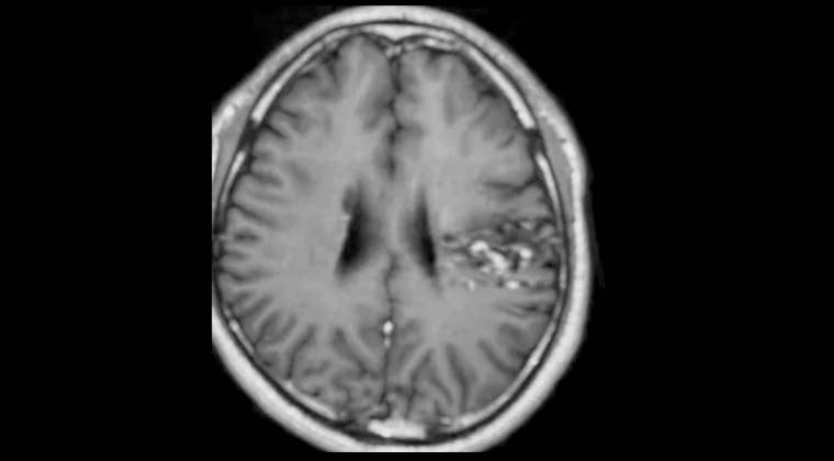
Figure 3: MRI follow-up on 2023-02-22 showing a significant reduction of the malformed vessels, seizures have resolved.
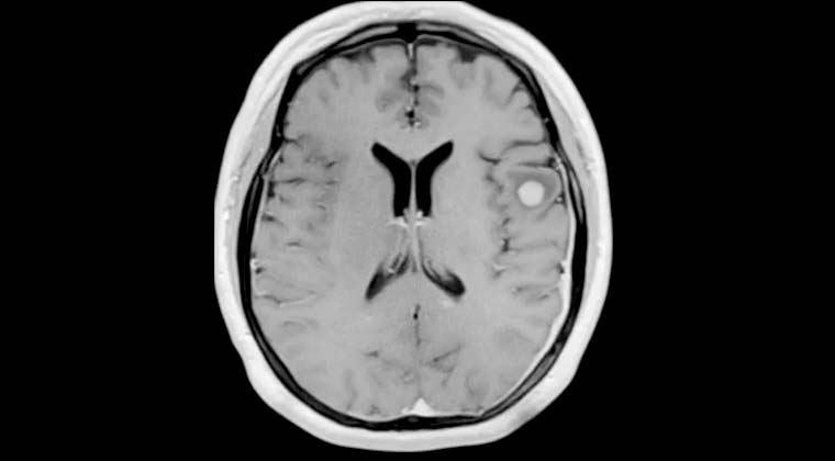
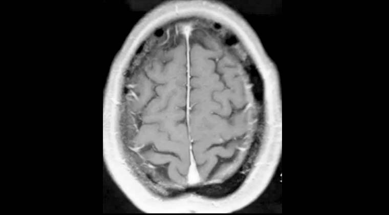
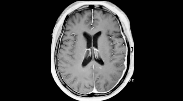
② Case 2:
Patient: Female, 56 years old
Chief complaint: weakness in the left limbs and difficulty walking for 2 weeks. Diagnosis: multiple brain metastases from lung cancer.
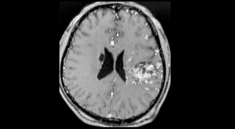
Figure 1: 2022‑02‑25 Gamma Knife treatment planning for the right frontal tumor with severe perilesional edema.
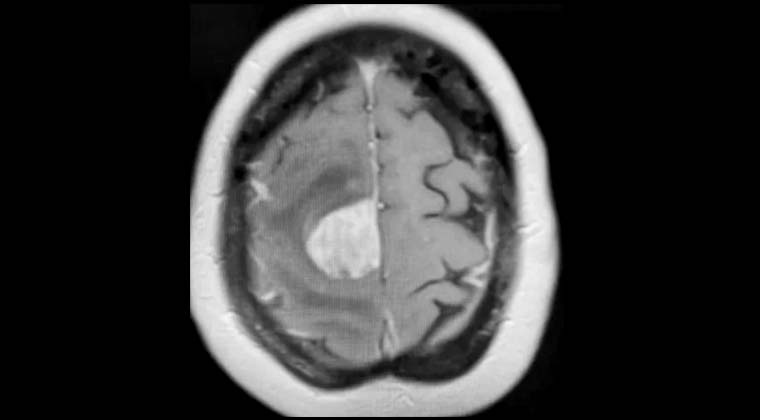
Figure 2: 2022‑02‑25 Gamma Knife treatment of the left temporal tumor performed simultaneously.
Figure 3: 2022‑07‑25, 5 months post-treatment, follow-up MRI showed the right frontal tumor had resolved and the patient was able to walk independently.
Figure 4: 2022-07-25, follow-up MRI shows the left temporal tumor has also disappeared.
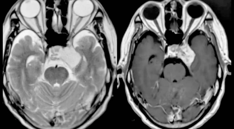
③ Case 3:
Patient: male, 65 years old
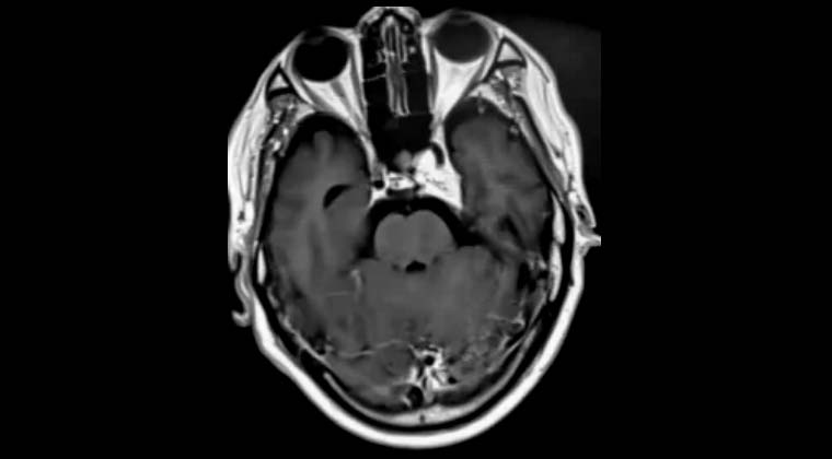
Cavernous hemangioma of the cavernous sinus
Figure 1: 10.06.2020 Gamma Knife treatment planning
Figure 2: 06.01.2021, 6 months after Gamma Knife treatment, the lesion significantly reduced
 + 7 (700) 356-49-30
 Address
 Email Form
 Language
 English

 + 7 (700) 356-49-30+ 86 13431079214+ 86 17688273501