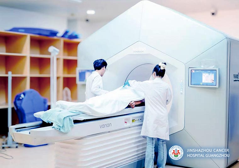
Halcyon Intelligent Linear Accelerator Radiotherapy
Applicable Situations
① Typical Scenarios Best Suited for Halcyon
• Patients mainly receiving conventional fractionated radiotherapy and intensity-modulated radiotherapy who require high-frequency imaging verification to improve setup consistency.
• Treatment scenarios involving complex anatomical regions such as the head and neck, thorax and abdomen, and pelvis, where accurate daily position correction is highly dependent.
• Patients whose thoracic or abdominal regions are significantly affected by respiratory motion, who have difficulty cooperating with breath-hold techniques, or who have reduced pulmonary function and wish to reduce treatment cooperation burden.
• Patients requiring complex immobilization with a high level of difficulty in reproducible positioning.
② Common Indications
• Head and neck tumors: nasopharyngeal carcinoma and other head and neck malignancies.
• Thoracic tumors: lung cancer, breast cancer, esophageal cancer.
• Abdominopelvic tumors: rectal cancer, prostate cancer.
• Bone tumors, intracranial tumors, and other types of malignancies.
Technical Advantages
① Faster and clearer three-dimensional imaging for a more comfortable treatment experience
• Equipped with a large-field imaging capability of 86 cm × 43 cm, providing broad coverage to meet the needs of comprehensive imaging for complex anatomical structures. Its wide field of view is particularly advantageous in pelvic radiotherapy, allowing clear visualization of the entire pelvic anatomy in a single scan, thereby reducing the time and radiation dose burden associated with repeated scanning for patients.
• HyperSight delivers near diagnostic-quality and simulation-grade imaging, providing more reliable image support for setup verification, target delineation, and dose optimization calculations. This allows better balance between target coverage and protection of normal tissues, making treatment safer and more reassuring.
• Compared with conventional iCBCT, scan time is reduced from 15 seconds to approximately 6 seconds. Patients do not need prolonged breath-holding, significantly reducing motion artifacts caused by respiration. This improves setup verification efficiency and comfort for motion-prone regions such as the chest and abdomen, lowering cooperation burden and breath-hold requirements and making the treatment process easier.
② SmART intelligent adaptive radiotherapy with rapid in-room adjustment for smoother treatment
With the combination of Halcyon and HyperSight, SmART adaptive plan modification can be performed, enabling more efficient plan adjustment and dose recalculation directly within the treatment room. This achieves the goals of "clear visualization, precise delivery, and rapid adjustment." Compared with traditional workflows that require treatment interruption, re-simulation, and subsequent return to treatment, this capability enhances treatment continuity and team efficiency, advancing precision radiotherapy from a "static plan" to a new stage of "dynamic response."
③ Efficient and standardized quality control for safer and more consistent treatment
• The dual-layer multileaf collimator (MLC) adopts a staggered double-layer design, which increases treatment speed while significantly reducing interleaf leakage. This allows more refined treatment planning while maintaining both efficiency and quality.
• Image-guided radiotherapy (IGRT) is deeply integrated into the treatment workflow, standardizing the sequence of "image acquisition, position correction, and irradiation" as routine practice. Each treatment is delivered on the basis of image verification and setup correction, improving consistency and safety between planned and delivered doses.
Why Choose Jinshazhou Hospital of Guangzhou University of Chinese Medicine?
Since introducing the Halcyon system in 2019, our hospital has treated more than ten thousand patients, covering tumors in all body regions, and has established a relatively mature quality control system and clinical pathway management framework. On September 15, 2025, our hospital completed the upgrade of the Halcyon linear accelerator to version 4.0 and integrated the advanced HyperSight image-guided technology, becoming the first radiotherapy center in mainland China to realize this Varian technology combination upgrade. This milestone further enhanced treatment precision, speed, and safety.
At present, HyperSight has been routinely applied to setup verification for tumors of the head and neck, thoracic and abdominal regions, and the pelvis. Relying on its high-quality image-guided capabilities, the team can perform more thorough setup verification and correction prior to treatment, improving the consistency between delivered irradiation and planned dose while better balancing target coverage and protection of normal tissues. Under appropriate indications and workflow conditions, high-quality imaging and more efficient plan adjustment can be completed directly in the treatment room, allowing therapy to better match the patient's physical condition at each session, helping shorten time spent in the treatment suite, reducing the time burden caused by repeated procedures, and improving the overall patient experience.
At the same time, our hospital advances one-stop international medical services with MDT as the core, bringing together multidisciplinary expertise from internal medicine, surgery, radiotherapy, interventional therapy, imaging, pathology, and nursing for collaborative decision-making and dynamic evaluation. Radiotherapy is incorporated into the overall pathway management of comprehensive cancer treatment. With Halcyon v4.0 equipped with HyperSight, our hospital has further enriched its options for high-quality image-guided and standardized workflow-based treatments. Looking ahead, we will continue to explore advanced treatment models such as adaptive radiotherapy and hypofractionated radiotherapy, providing more individualized, efficient, and safer treatment options for a wider range of patients, and steadily translating technological advantages into tangible improvements in treatment quality and patient experience.
Technical Definition
Halcyon v4.0 is an integrated, fully digital intelligent linear accelerator radiotherapy platform in which image guidance is embedded throughout every treatment session. Equipped with the advanced HyperSight imaging guidance technology, the system leverages coordinated hardware and algorithmic optimization to achieve large-field-of-view, high-quality, and rapid three-dimensional imaging. This provides a reliable basis for patient positioning verification, target delineation, and dose optimization, while supporting the SmART intelligent adaptive radiotherapy workflow.
Mechanism of Action / Working Principle
Halcyon v4.0 generates high-energy photon beams via a linear accelerator to deliver precise radiation to tumor targets, causing DNA damage in tumor cells and thereby inhibiting their growth and division. Before treatment, three-dimensional image guidance is used for positioning verification and alignment correction, reducing the risk of deviation. HyperSight provides large-field-of-view, high-quality, rapid three-dimensional imaging (86 cm × 43 cm, approximately 6 seconds acquisition), minimizing anatomical truncation and motion artifacts. Under applicable indications and workflow conditions, this imaging can be used to detect anatomical changes and implement SmART adaptive plan adjustments, making radiation delivery better aligned with the patient's current anatomical status.
Treatment Procedure
① Assessment and Treatment Planning: Evaluate the patient using pathological and imaging data (CT/MRI/PET, etc.); involve MDT if necessary to define treatment goals and organ protection principles.
② Simulation and Positioning: Secure patient positioning (e.g., mask/body mold) and acquire reference imaging to establish a baseline for planning.
③ Contouring and Plan Verification: Physicians delineate the target area and organs at risk, physicists complete dose design and optimization, and the team reviews and confirms the plan for execution.
④ Daily Patient Positioning: Patients arrive according to schedule, are positioned, and identity is verified.
⑤ Image-Guided Correction: Prior to each session, HyperSight acquires three-dimensional images (iCBCT) for positioning verification and alignment correction before irradiation, enhancing accuracy and safety.
⑥ Adaptive Adjustment if Needed: Under applicable indications and workflow conditions, if imaging shows anatomical changes affecting dose distribution, SmART adaptive plan adjustments can be made and verified before treatment.
⑦ Course Management and Follow-Up: Complete the treatment course fraction by fraction, monitor patient response, provide supportive care, and schedule follow-up visits to evaluate efficacy and recovery.
Frequently Asked Questions
① Does the faster iCBCT scan mean patients still need to hold their breath?
Answer: HyperSight acquires images in approximately 6 seconds, reducing the need for prolonged breath-holding. This generally helps minimize respiratory-related artifacts and improve patient compliance. Whether breath-hold is required depends on the treatment site and physician's instructions.
② When is SmART adaptive radiotherapy used?
Answer: If the imaging during a session indicates changes in the target or surrounding tissues that may affect dose distribution, the team evaluates whether to enter the adaptive workflow. Under applicable indications and workflow conditions, target/organ adjustments and dose recalculation can be performed to better match the current anatomical state.
③ If a planning CT has already been done, why is 3D imaging needed on the treatment day?
Answer: Planning images establish a baseline for the treatment plan; images taken on the treatment day verify the patient's position and current anatomical state. Together, they ensure consistency between the planned and delivered dose while balancing target coverage and normal tissue protection.
④ Is hospitalization required?
Answer: Most patients can complete treatment on an outpatient basis, attending scheduled sessions. If closer nursing support or combined treatments are needed, the team will assess and recommend the most suitable option (outpatient or short-term hospitalization).
⑤ What precautions should patients take during treatment?
Answer: Patients should cooperate with the medical team for positioning and planning procedures, maintain a stable treatment posture, and follow medical staff guidance. Any discomfort should be reported promptly. After radiotherapy, attention should be given to skin care, dietary adjustments, and regular follow-up to evaluate treatment efficacy.
Typical Case
① Case 1:
Adolescent male, 13 years old
Pineal germ cell tumor
One month ago, the patient developed headaches at school without obvious cause. Initially ignored, the headaches progressively worsened and were accompanied by unsteady walking. The family sought medical attention. Cranial MRI revealed a pineal mass with obstructive hydrocephalus. Blood tests showed mildly elevated HCG and AFP within normal limits. Clinical diagnosis: Pure pineal germ cell tumor.
Precision radiotherapy was administered at 9 Gy/5 fractions. The pineal tumor significantly reduced in size (red arrow in image indicates tumor), hydrocephalus improved, headaches largely resolved, and unsteady walking improved. Subsequent treatment followed standard chemoradiotherapy for intracranial germ cell tumors.
Before Treatment
5 fractions of radiotherapy
② Case 2:
Male patient, 49 years old
Diffuse large B-cell lymphoma
On 2021-03-03, prior to radiotherapy, a plain and contrast-enhanced cranial MRI was performed:
Lesion in the body of the corpus callosum extending to the parietal lobe, consistent with lymphoma.
On 2021-03-11, cranial lesion radiotherapy was performed using 6MV X-rays, with simultaneous integrated boost: PTV1: 40 Gy / 2 Gy / 20 fractions; PTV2: 36 Gy / 1.8 Gy / 20 fractions; 5 fractions per week
Follow-up MRI on 2021-04-07 showed post-treatment changes consistent with lymphoma, with intracranial lesions smaller than before.
Before Treatment:
After treatment:
③ Case 3:
Female patient, 59 years old
Bladder cancer
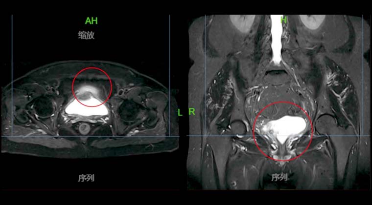
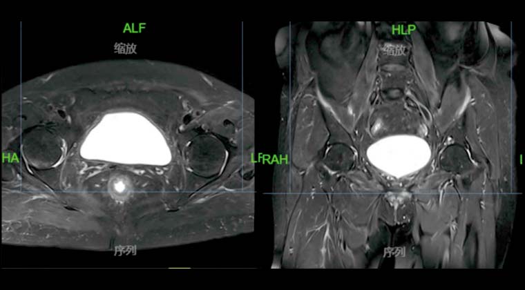
On 2021-03-29, PET/CT examination showed multiple lesions on the anterior, posterior, and base walls of the bladder, considered tumor recurrence.
Concurrent chemoradiotherapy was administered (single-agent cisplatin 20 mg/m² weekly) with pelvic VMAT 45 Gy, with local bladder boost of 20 Gy, under IGRT guidance.
One month after radiotherapy, pelvic MRI follow-up showed no residual tumor or signs of recurrence in the bladder.
Before Treatment:
After treatment:
 + 7 (700) 356-49-30
 Address
 Email Form
 Language
 English

 + 7 (700) 356-49-30+ 86 13431079214+ 86 17688273501