Indications
Radiofrequency ablation is indicated for the local treatment of various solid tumors, particularly in patients who are not candidates for surgery or for whom surgery is not advisable. Common indications include:
① Primary tumors:
Early-stage hepatocellular carcinoma that cannot be surgically resected or is unsuitable for surgery, especially in patients with small tumor size and a limited number of lesions;
② Recurrent tumors:
Local recurrent lesions after surgery or following multiple prior treatments, serving as an important option for repeat local disease control;
③ Metastatic tumors:
Liver metastases or other localized metastatic lesions originating from gastrointestinal, breast, lung, uterine, and other primary tumors;
④ Palliative and combination therapy:
In patients with intermediate to advanced-stage tumors, radiofrequency ablation may be used to alleviate symptoms, control tumor burden, or as an integral component in combination with interventional therapy, radiotherapy, or systemic treatment.
Core Advantages
① Minimally invasive and safe
The procedure is performed via percutaneous puncture without open surgery, resulting in minimal trauma, rapid recovery, and lower physical burden for the patient.
② Precise ablation
Under real-time ultrasound or CT guidance, energy is accurately delivered to the tumor itself, minimizing damage to surrounding normal tissues.
③ Broad patient applicability
Particularly suitable for patients with liver cirrhosis, multiple comorbidities, or poor tolerance for surgical procedures.
④ Repeatable treatment
Can be performed multiple times under appropriate conditions, providing ongoing treatment options for recurrent or multifocal lesions.
⑤ Flexible combination therapy
Can be combined with interventional embolization, systemic therapy, radiotherapy, and other modalities to enhance overall treatment efficacy.
Why Choose Jinshazhou Hospital?
Our hospital has accumulated extensive clinical experience in radiofrequency ablation for tumor treatment and has established a therapeutic system characterized by image guidance and minimally invasive precision. Supported by comprehensive imaging platforms (ultrasound, CT, PET-CT), lesions can be accurately assessed and guided in real time, ensuring adequate ablation coverage with controllable safety.
Radiofrequency ablation is planned and implemented following joint evaluation by an experienced multidisciplinary team (MDT). Treatment strategies are individualized based on tumor type, location, and the patient's overall condition, and can be coordinated with interventional therapies, radiotherapy, systemic treatment, and traditional Chinese medicine.
While prioritizing safety, this approach helps achieve improved local tumor control and better quality of life for patients.
Technical Definition
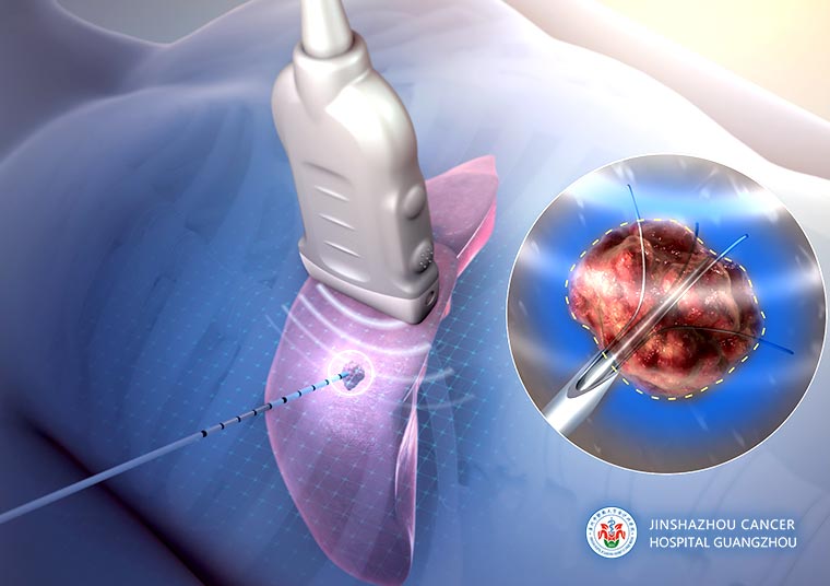
Radiofrequency ablation (RFA) is a well-established and classical minimally invasive tumor ablation technique. Under real-time imaging guidance, a radiofrequency electrode needle is precisely inserted into the tumor, where thermal energy generated by radiofrequency current rapidly increases local tissue temperature, resulting in coagulative necrosis and loss of viability of tumor cells.
This technique does not require open surgery, is associated with minimal trauma and rapid recovery, and has been widely applied in the treatment of various solid tumors, including those of the liver, lung, kidney, adrenal gland, and bone metastases. It represents an important component of modern minimally invasive oncologic therapy.
Working Principle / Mechanism
Radiofrequency ablation uses a radiofrequency generator to produce high-frequency alternating current, which is delivered to the tumor tissue through an electrode needle, causing rapid ionic vibration and friction and generating sustained heat.
When the tissue temperature rises above 60 °C (typically reaching 90–100 °C), intracellular proteins undergo denaturation and cell membranes are disrupted, ultimately resulting in irreversible coagulative necrosis. At the same time, small blood vessels surrounding the tumor are thermally coagulated, reducing blood supply and expanding the ablation zone.
Through one or multiple ablation sessions, tumor tissue can be progressively reduced and inactivated. The entire procedure is performed under real-time imaging guidance such as ultrasound or CT, ensuring precision and safety of the treatment.
Treatment Procedure
① Pre-procedural assessment: Imaging studies are performed to evaluate the tumor's location, size, number, and its relationship to critical structures, together with a comprehensive assessment of the patient's general condition and organ function.
② Imaging-guided localization: The lesion is precisely localized under ultrasound or CT guidance.
③ Electrode needle insertion: The radiofrequency electrode needle is inserted into the tumor via percutaneous puncture (laparoscopic or open approaches may be used if necessary).
④ Radiofrequency ablation: Radiofrequency energy is delivered, generating high temperatures within the tumor tissue and inducing coagulative necrosis.
⑤ Post-procedural observation and follow-up: Short-term observation is conducted after the procedure, followed by regular follow-up with contrast-enhanced CT or MRI to assess the ablation effect.
Safety and Precautions
Radiofrequency ablation is generally considered safe; however, strict selection of indications and standardized procedures are essential.
Before treatment, thorough evaluation of liver function, tumor location, and anatomical relationships with surrounding critical organs is required to determine an appropriate puncture pathway. Transient fever or localized pain may occur after the procedure and usually resolves spontaneously. In rare cases, complications such as infection, bleeding, or irritation of adjacent organs may occur; therefore, the procedure should be performed in medical centers equipped with comprehensive imaging guidance and an experienced multidisciplinary team.
Through standardized assessment and precise technique, treatment safety and effectiveness can be maximized.
Frequently Asked Questions
① How soon can normal daily life be resumed after treatment?
Most patients recover relatively quickly after radiofrequency ablation and can usually resume daily activities gradually within 1–2 weeks after the procedure. Strenuous exercise and heavy physical labor should be avoided in the short term, and follow-up examinations should be performed as advised by the physician.
② Is repeated treatment necessary?
Whether repeated treatment is required depends on the size, number, and location of the tumor. Some patients may need multiple sessions of radiofrequency ablation or include it as part of a comprehensive treatment plan to achieve better local control.
③ How soon can the treatment effect be observed?
Immediate effect: Within several days to one week after treatment, the local tumor tissue gradually undergoes coagulative necrosis, and some patients may notice a reduction in local discomfort.
Final effect: Definitive evaluation is usually performed 1–3 months later using contrast-enhanced CT or MRI, based on which residual disease or recurrence is assessed.
Representative Case
Before ablation
After ablation
 + 7 (700) 356-49-30
 Address
 Email Form
 Language
 English

 + 7 (700) 356-49-30+ 86 13431079214+ 86 17688273501