Common Indications
① Malignant tumors
Liver cancer, pancreatic cancer, retroperitoneal metastatic tumors, bladder cancer, renal cancer, rectal cancer, prostate cancer, ovarian cancer, endometrial cancer, abdominopelvic tumors, breast cancer, leiomyosarcoma, liposarcoma, soft tissue sarcoma, etc.
② Benign tumors
Uterine fibroids, adenomyosis, breast fibroadenoma, benign prostatic hyperplasia, benign soft tissue tumors, etc.
Core Advantages
HIFU is characterized by non-invasiveness, controllability, conformal coverage, and repeatable evaluation, providing a local treatment option for selected patients with tumors and benign diseases.
① Truly extracorporeal and non-invasive: no puncture, no incision
HIFU delivers ultrasound energy from outside the body and focuses it within the target lesion to achieve treatment. The entire procedure requires no percutaneous puncture or instrument insertion, avoiding incision-related risks such as bleeding and infection and helping to shorten the recovery period.
② High energy density at the "focal point": rapid heating with concentrated thermal effect
Ultrasound energy forms a millimeter-scale focal point within the target area, where local temperatures can rapidly rise to approximately 70--100 °C, achieving fast thermal coagulation and ablation. As the energy passes through the skin and surrounding tissues, it is relatively dispersed, which helps reduce heating of non-target areas.
③ Organ morphology and function preservation--oriented: suitable for patients requiring functional preservation
HIFU energy primarily acts at the focal point within the target area, while surrounding tissues are usually outside the focus. Under appropriate indications and standardized operation, it helps achieve local control while minimizing impact on overall organ morphology and function.
④ Real-time imaging monitoring + computer-precise control: visualized, adjustable, and assessable
The system integrates imaging localization with computer control, enabling real-time monitoring of the target area and energy deposition during treatment. Layered planning can be performed according to tumor morphology, with stepwise scanning from "point--line--plane--volume" to achieve refined target coverage. Parameters can be dynamically adjusted when needed, enhancing process controllability and safety margin management.
⑤ No use of ionizing radiation: a physical modality within comprehensive treatment strategies
HIFU does not involve X-ray exposure or radioactive sources. As a local physical treatment modality, it can be considered---after MDT evaluation---in combination with radiotherapy, chemotherapy, and other comprehensive approaches, meeting strategic needs across different stages of treatment.
Why choose Jinshazhou Hospital?
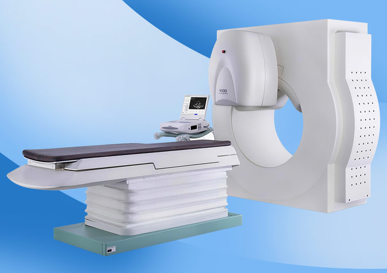
Our hospital positions HIFU as one of the "truly non-invasive local physical treatment" modalities, focusing on the needs of patients who seek reduced trauma and emphasize preservation of organ function. The hospital is equipped with a third-generation HIFU system, offering clear platform advantages in treatment experience and operational flexibility: the overhead treatment design allows for better patient comfort and cooperation; more favorable temperature management along the acoustic entry pathway helps reduce the risk of skin thermal reactions; dual-focus transducers can be flexibly combined according to lesion location to optimize the acoustic path and avoid obstacles such as ribs; the treatment head supports multi-directional rotation, enabling conformal scanning that progresses "from point to line, from line to plane, and from plane to volume," better matching complex target areas.
In clinical practice, the hospital emphasizes a standardized HIFU workflow that is "visualized, plannable, and assessable." Before treatment, ultrasound imaging is acquired and three-dimensional reconstruction is performed to plan scanning layers. During treatment, focal position and energy delivery are dynamically adjusted under real-time ultrasound monitoring and computer control. After treatment, imaging follow-up is used to evaluate response, creating conditions for subsequent integration into comprehensive treatment strategies and long-term management.
Through the combination of "advanced equipment platform advantages + standardized process control," our hospital provides patients with a more stable and controllable non-invasive treatment option.
Technical Definition
HIFU, or High-Intensity Focused Ultrasound, is commonly known in Chinese as "Haifu" due to the phonetic similarity of the English abbreviation. It has been referred to by experts both in China and internationally as a "new non-invasive treatment technology of the 21st century" and a "green tumor treatment technology."
HIFU is a non-invasive local treatment modality in which ultrasound energy is emitted extracorporeally and focused within the body. Under image guidance and computer control, incision-free local thermal ablation is applied to the target area. By concentrating ultrasound energy on the lesion, the tissue at the focal point rapidly increases in temperature and undergoes coagulative necrosis, thereby achieving local ablation and control of the target area. Its key characteristics are the absence of incisions and its applicability to the local treatment of various solid tumors and certain benign diseases.
Working Principle / Mechanism
HIFU (High-Intensity Focused Ultrasound) makes effective use of the favorable directionality, penetration, and focusability of ultrasound waves in biological tissues.
During treatment, an external ultrasound transducer emits and focuses ultrasound energy. Under imaging monitoring and computer control, the acoustic energy is precisely concentrated within the intratumoral target area to form a focal zone (approximately Φ3 × 3 × 8 mm). When energy is highly concentrated at the focus, the temperature of the tumor tissue in the target area can rapidly rise to approximately 70--100 °C, leading to protein denaturation, coagulation, and necrosis at the focal point; cavitation effects and related biological effects may also occur, further enhancing local tumor inactivation.
Throughout the procedure, monitoring ultrasound acquires tumor images and performs three-dimensional reconstruction. The target area is planned layer by layer according to tumor morphology, after which the focal point is scanned sequentially in a "point-to-line, line-to-plane, plane-to-volume" manner, progressing from the first layer to the last to complete thermal coagulative ablation of the entire tumor volume. Because the energy is mainly concentrated at the focal point, surrounding normal tissues are usually outside the focus; with standardized procedures and appropriate indication selection, the impact on non-target tissues can be minimized.
Treatment Workflow
① Pre-treatment evaluation and examinations
Routine ultrasound examination and relevant imaging assessment are performed, with clinical exclusion of infection. The treatment pathway and lesion location, size, and relationship to surrounding organs are evaluated, and the presence of metal or other factors that may interfere with treatment at the target area is excluded.
② Pre-treatment preparation
Bowel preparation: if the mass is located in the abdominopelvic region, high-protein foods are avoided on the day before treatment; fasting is required for 10 hours before treatment; oral laxatives and cleansing enemas may be used when necessary to reduce intestinal gas interference; a light, low-residue diet is recommended for 3 days prior to treatment.
Skin preparation: generally not required; hair removal at the treatment area may be performed if necessary.
Bladder preparation: if the tumor is located in the bladder, prostate, or other lower abdominal regions, bladder filling may be carried out as instructed by the physician.
③ Positioning and localization
The patient lies supine on the treatment table (position may be adjusted as needed) with full exposure of the treatment area. Under ultrasound monitoring and computer-assisted positioning control, tumor images are acquired, three-dimensional reconstruction is performed, and scanning layers are planned.
④ Focused irradiation and layer-by-layer scanning
The external ultrasound transducer delivers focused ultrasound. According to tumor morphology, the focal point is scanned sequentially in a "point-to-line, line-to-plane, plane-to-volume" manner, progressing from the first layer to the last to achieve complete coverage of the target area.
⑤ Post-treatment observation and follow-up
Pain, body temperature, and skin reactions are monitored. Follow-up examinations and imaging assessments are performed as prescribed to evaluate the treatment response.
Safety and Precautions
① Pre-treatment considerations
• Clinical exclusion of infection; assessment of the acoustic window conditions and critical structures surrounding the lesion; avoidance of metal or other interfering factors at the treatment site.
• For abdominopelvic lesions, bowel preparation is often required to reduce interference from intestinal gas; for lower abdominal lesions, bladder preparation is performed as needed.
• In cases of severe cardiac, pulmonary, or renal disease, or inability to cooperate with treatment, suitability for the procedure should be comprehensively evaluated by the physician.
② Common post-treatment adverse reactions and management
• Pain: relatively common, especially during treatment of superficial tumors. Analgesics may be administered when necessary, with careful monitoring of the skin to prevent thermal injury.
• Fever: relatively common, usually caused by absorption of necrotic tumor tissue after treatment; it generally resolves spontaneously within 48 hours, or can be managed with symptomatic antipyretic treatment.
• Skin erythema, blistering, burns: occasional; after treatment, cold compresses with ice packs at 0--4 °C for 10--30 minutes may be applied; if necessary, burn ointments, local dressing changes, and antibacterial agents can be used for symptomatic management.
• Nerve injury: very rare; neurotrophic agents may be administered, with recovery usually occurring within approximately two weeks after the procedure.
Frequently Asked Questions
① Does HIFU require surgery or puncture?
No. HIFU is a non-invasive treatment in which extracorporeal ultrasound energy is focused on the target area inside the body under ultrasound guidance to complete the therapy.
② Is the treatment painful? Is anesthesia required?
Some patients may experience pain during or after treatment, which is more common with superficial tumors. The need for anesthesia or analgesia depends on the lesion location, treatment extent, and individual tolerance. Symptomatic pain relief can be provided when necessary.
③ Why do some patients require bowel preparation or bladder preparation?
Pelvic and abdominal masses may be affected by intestinal gas. To reduce interference, dietary control, cleansing enemas, and other bowel preparations may be performed before treatment as prescribed. For lower abdominal lesions (such as those in the bladder or prostate region), bladder filling may sometimes be required to facilitate treatment localization and optimize the acoustic pathway.
 + 7 (700) 356-49-30
 Address
 Email Form
 Language
 English

 + 7 (700) 356-49-30+ 86 13431079214+ 86 17688273501