Large Bore CT Simulation
Applicable Situations
① Common Scenarios
• Lesions with complex shapes or located near critical organs, requiring multi-angle or rotational irradiation.
• Sites significantly affected by physiological movements such as respiration (e.g., thoracic or abdominal regions).
• Patients or sites requiring positioning devices or special positioning.
• Large body habitus (e.g., obesity) or areas needing wide surface coverage / large field of view.
• Areas with dental metal, orthopedic implants, or regions at high risk of artifacts.
② Common indications
Gliomas, pituitary tumors, brainstem tumors, metastases, maxillary sinus cancer, nasopharyngeal carcinoma, laryngeal cancer, lung cancer, breast cancer, liver cancer, pancreatic cancer, prostate cancer, rectal cancer, bone and soft tissue tumors, etc.
Core Advantages
① 4D Dynamic Imaging: Reduces motion effects, improves localization reliability
The large-bore CT system can perform respiratory-gated 4D imaging to analyze tumor and organ motion throughout the breathing cycle. This provides information on motion range and patterns for radiotherapy planning, reduces localization uncertainties caused by motion, and minimizes repeated adjustments due to respiratory effects.
② Clearer Images: Better boundary delineation and more accurate contouring
The large-bore CT system offers high spatial resolution and excellent gray-white tissue differentiation, clearly showing tumor boundaries and their relationship with surrounding organs. This provides a reliable basis for target delineation and allows treatment planning to be more precise.
③ Larger Aperture and Wider Field of View: Easier positioning, more comfortable experience
The 80 cm large aperture and wide field of view facilitate patient positioning and special postures. It is especially suitable for larger patients or those requiring extensive coverage.
④ Dual-Energy Imaging: Fewer artifacts, clear images even in complex cases
For patients with dental metal, orthopedic implants, or areas prone to artifacts, dual-energy scanning improves contrast and reduces metal artifact interference, minimizing impact on contouring and planning. Complex cases can still obtain stable, high-quality localization images.
⑤ Stable Under High Load: Smoother workflow, less waiting
Equipped with a STRATON tube with zero megavoltage and enhanced cooling, the tube can handle up to 50 MHU (about six times that of conventional large-bore CT). High-load scans are less likely to trigger cooling delays, ensuring a smoother workflow.
⑥ Low-Dose Technology: Minimizes radiation while maintaining image quality
Incorporates multiple low-dose protocols and real-time 4D dose modulation to reduce scanning dose as much as possible while ensuring image quality, decreasing unnecessary radiation exposure to patients.
⑦ Consistent Electron Density: More accurate planning, fewer errors
Direct Density™ "electron density normalization" technology expands CT voltage selection, providing flexible parameter options. It maintains planning accuracy, improves efficiency, and reduces errors from manual settings.
Why Choose Jinshazhou Hospital?
Compared with conventional radiotherapy, smart precision radiotherapy relies heavily on accurate localization. In our radiotherapy workflow, CT simulation is the critical first step: imaging is acquired in the treatment position to give doctors a clear understanding of tumor location and its relationship with surrounding organs at risk, providing a reliable basis for target contouring, plan design, and treatment delivery. To establish a stable foundation for this step, our hospital was the first in China to introduce the Siemens dual-energy photon 4D large-bore helical CT simulator SOMATOM Confidence, marking a major milestone in the development of our precision radiotherapy platform.
Based on this, we have developed a well-established workflow covering imaging acquisition, patient positioning, respiratory management, and post-processing, seamlessly integrating with subsequent radiotherapy platforms. For challenging scenarios, such as tumors affected by motion, special positioning requirements, or metal-induced artifacts, this system provides more stable localization support, facilitating precise planning and smooth treatment delivery.
Over the years, we have continuously optimized team coordination and operational procedures for simulation and localization, emphasizing process standardization and end-to-end management: imaging results are reviewed and quality-controlled before treatment proceeds; within an MDT framework, the timing and role of radiotherapy in the overall treatment plan are clearly defined, coordinated with surgery, interventional procedures, and systemic therapy; during treatment, follow-up imaging and plan reassessment are performed promptly to refine strategies, creating a continuous management loop.
For patients, our focus goes beyond advanced equipment. We invest in standardized workflows, quality assurance, and multidisciplinary collaboration to ensure that each localization is reliable, each treatment is safe, and the overall patient experience is smooth.
Technical Definition
The large-bore CT simulator is a key device in the precision radiotherapy workflow. It acquires and reconstructs imaging in the treatment position, providing critical imaging data for target delineation, dose calculation, and treatment planning. This enhances the accuracy and feasibility of radiotherapy plans while minimizing unnecessary exposure to surrounding healthy tissues. Our hospital is equipped with the Siemens SOMATOM Confidence dual-energy photon 4D large-bore helical CT simulator, which supports rapid image acquisition for various anatomical sites and patient positions, providing a reliable imaging foundation for radiotherapy planning.
Mechanism of Action / Working Principle
① Dual-energy scanning: The device acquires data using X-rays at two different energy levels. Because different tissues attenuate X-rays differently at each energy, the system can better distinguish tissue composition and generate additional image information for analysis.
② Four-dimensional (4D) imaging: "Four-dimensional" refers to adding the time dimension to three-dimensional space. By continuously acquiring images with respiratory gating, images at different phases of the breathing cycle are reconstructed separately to observe tumor/organ motion during respiration.
③ Large-bore helical scanning: During scanning, the X-ray tube and detectors rotate continuously around the patient while the examination table moves steadily along the body's long axis, forming a helical acquisition trajectory. This allows rapid acquisition of large-volume thin-slice images.
④ Photon detection: The system uses StellarRT photon detectors to receive X-ray signals and convert them into electrical signals, which are then digitized to form imaging data. These detectors minimize electronic noise and signal loss, improving spatial resolution and providing a reliable data foundation for dual-energy and other clinical applications.
⑤ Data processing and image reconstruction: Raw data are corrected, reconstructed, and post-processed to generate multiplanar and three-dimensional images, serving as the fundamental imaging data for radiotherapy localization and treatment planning.
Treatment Procedure
① Patient preparation and information verification: After arrival at the simulation room, medical staff verify the patient's identity and the target site for simulation, and explain the examination procedure and cooperation requirements. If special preparation is required (such as contrast enhancement or fasting), the medical team will inform the patient in advance and assist with arrangements.
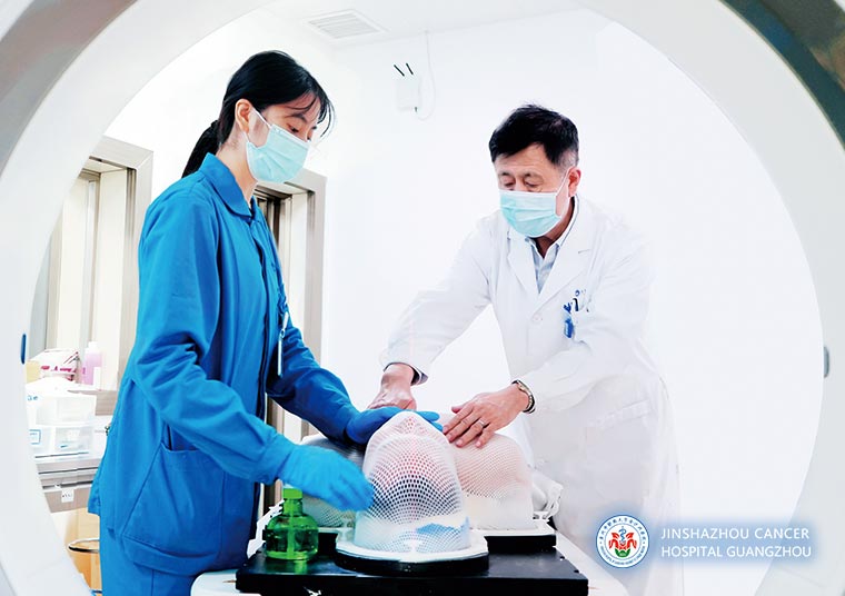
② Positioning and immobilization: According to the simulation site, the patient is placed in an appropriate position (supine, prone, arms raised, etc.). Immobilization devices such as thermoplastic masks, headrests, and cushions are used to fix the patient's position and relevant body parts, ensuring positional stability during scanning and enabling accurate repositioning in subsequent treatments.
③ Respiratory coordination training: For sites significantly affected by respiration, such as the chest and abdomen, the patient may undergo brief breathing training. If respiratory gating is used, the patient follows the prompts to complete breathing coordination.
④ Simulation scanning: A low-dose scout scan is first performed to obtain preliminary images and determine the scan range and starting position. This is followed by the formal simulation scan (with dual-energy and/or 4D acquisition when necessary). During scanning, medical staff monitor the patient via observation and intercom. The patient is instructed to remain still; in case of discomfort or emergency, the patient may raise a hand or inform staff via the intercom.
⑤ Marking and documentation: After scanning, reference marks are placed on the patient's skin (drawing lines or applying markers), and the positioning and immobilization methods are documented to facilitate accurate repositioning during subsequent treatments.
⑥ Treatment planning and transition to therapy: The simulation images are transferred to the radiotherapy planning system. The radiotherapy team performs image evaluation and completes treatment plan generation and verification. After plan confirmation, the patient proceeds to subsequent radiotherapy steps according to the schedule.
Frequently Asked Questions
① How long does CT simulation take? Is it painful?
The scan itself is usually quick. Most of the time is spent on patient positioning and immobilization, especially when special positions or thermoplastic masks are required. The exact duration depends on on-site arrangements. CT simulation is generally not painful.
② What is contrast-enhanced scanning? When is it needed?
Contrast-enhanced scanning usually refers to intravenous injection of an iodine-based contrast agent to better delineate blood vessels, tumors, and their boundaries with surrounding structures, making it easier for physicians to identify key anatomy on simulation images. Whether contrast is required is determined by the physician based on the simulation site and planning needs.
③ What should be noted on the day of examination?
It is recommended to wear loose clothing and remove metal objects as instructed by medical staff. During scanning, remain still as directed. If respiratory coordination is required, follow the breathing instructions. If anxiety, coughing, or discomfort occurs, inform the medical staff promptly so they can adjust the process or provide further guidance.
④ Is the radiation dose higher with 4D CT simulation?
The radiation dose depends on the examined site and motion-related factors. To record information across different respiratory phases, 4D CT simulation involves more extensive data acquisition, and the dose may be higher than that of conventional CT simulation. The radiotherapy team will use low-dose strategies and dose modulation techniques whenever possible while meeting planning requirements to reduce unnecessary radiation exposure.
⑤ Is CT simulation still required if another CT has already been performed?
Even if a diagnostic CT has been performed recently, radiotherapy usually still requires a dedicated CT simulation, because it is acquired in the treatment position and is directly linked to radiotherapy planning and subsequent patient repositioning, ensuring accurate treatment delivery.
 + 7 (700) 356-49-30
 Address
 Email Form
 Language
 English

 + 7 (700) 356-49-30+ 86 13431079214+ 86 17688273501